Hip And Upper Thigh Anatomy / Like the forearm, the upper leg, or thigh, has a dense arrangement of many muscles.. The joint recess extends from the acetabulum over the femur to the level of the intertrochanteric line, just beyond the femoral neck. The bones of the hip include the femur, the ilium, the ischium, and the pubis. If the pain is located in the groin or in the thigh, this is known as psoas bursitis, which affects the muscle that connects the femur to the lumbar vertebrae. The femoral triangle is made up of the sartorius (laterally), the inguinal ligament (superiorly) and the adductor longus (medially) and is found at the upper thigh. The six hip adductor muscles are all located in the adductor or medial compartment of the thigh and all mainly adduct the thigh at the hip joint.
The hip joint is made up of two bones: The thigh bone or femur and the pelvis join to form the hip joint. It is also referred to as a ball and socket joint and is surrounded by muscles, ligaments, and tendons. • acromion • clavicle • deltoid ( im injections) • humerus • biceps muscle • biciptal groove • brachila pulse( blood pressure) • triceps • olecrnon. This nerve runs along the.
Femur Wikipedia from upload.wikimedia.org It can resist forces of 1,800 to 2,500 pounds, so it. The hip muscles are going to be slip into hip muscles and gluteal muscles. These three pelvic bones form the acetabulum, a deep socket where the top of the thigh bone (the ball) fits into the socket. The hip itself is a ball and socket joint, much like the shoulder.the structures necessary to create this joint are the socket, the joint capsule, muscle, ligaments, and the neck. The hip is formed where the thigh bone (femur) connects with the three bones of the pelvis: The ilium, the pubis (pubic bone) and the ischium. What movements does it control? Understanding the anatomy of the hip is essential for diagnosing its pathology.
The hip is formed where the thigh bone (femur) connects with the three bones of the pelvis:
The hip adductors shape the surface anatomy of the medial thigh. Sartorius muscle anatomy page has origin, insertion, innervation, and blood supply information. Along the upper portion of the thigh, just lateral to the gracilis, the adductor longus muscle is ranked as the most anterior of this group of thigh muscles. The hip joint receives innervation from all three nerves. The hip joint consists of two main parts: Hip and thigh anatomy the hip joint is a synovial articulation between the acetabulum of the pelvis and the proximal femur. Abductors are located on the upper portion of the outside of your thighs and hips, anchoring above on the pelvis, and below at various points on your outside thigh. It is very thin behind and. These muscles can be grouped based upon their location and function. The many muscles of the hip provide movement, strength, and stability to the hip joint and the bones of the hip and thigh. By adulthood, these three bones are completely fused and the pelvis is effectively a single bone. In some severe cases, physical. A collection of anatomy notes covering the key anatomy concepts that medical students need to learn.
Abductors are located on the upper portion of the outside of your thighs and hips, anchoring above on the pelvis, and below at various points on your outside thigh. The many muscles of the hip provide movement, strength, and stability to the hip joint and the bones of the hip and thigh. It is very thin behind and. It has branches that innervate the anterior thigh muscles and the hip joint. These are gracilis, pectineus, adductor longus, adductor brevis, adductor magnus, and adductor minimus muscles.
Part 4 Anatomies Of The Lower Limbs The Knee Thigh Hip And Groin Ppt Download from slideplayer.com The thigh muscles are divided into three compartments: The thigh is the region between the hip and knee joints. What movements does it control? These three pelvic bones form the acetabulum, a deep socket where the top of the thigh bone (the ball) fits into the socket. • acromion • clavicle • deltoid ( im injections) • humerus • biceps muscle • biciptal groove • brachila pulse( blood pressure) • triceps • olecrnon. In some severe cases, physical. It is also referred to as a ball and socket joint and is surrounded by muscles, ligaments, and tendons. Bony anatomy • acetabulum, femoral head and neck, greater and lesser trochanter, and femoral…
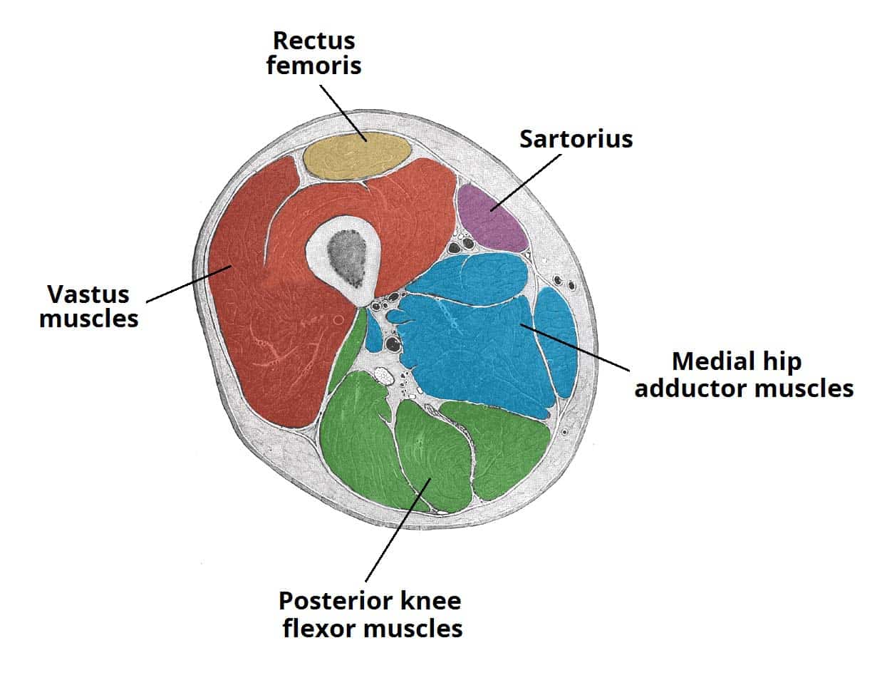
On the anterior side, the most prominent of the muscles are the sartorius muscle and the four muscles that make up quadriceps muscle group (the quads.)
Hip and thigh anatomy the hip joint is a synovial articulation between the acetabulum of the pelvis and the proximal femur. Bony anatomy • acetabulum, femoral head and neck, greater and lesser trochanter, and femoral… The hip joint is made up of two bones: Sartorius muscle anatomy page has origin, insertion, innervation, and blood supply information. By adulthood, these three bones are completely fused and the pelvis is effectively a single bone. Bf sh, lh, biceps femoris short head, long head; The hip joint is one of the most flexible joints in the entire human body. Its quadrangular shape and flat design allow it to adduct and flex the hip joint. The joint recess extends from the acetabulum over the femur to the level of the intertrochanteric line, just beyond the femoral neck. The gluteus maximus is the large muscle of the buttock. It is also referred to as a ball and socket joint and is surrounded by muscles, ligaments, and tendons. It is very thin behind and. Along the upper portion of the thigh, just lateral to the gracilis, the adductor longus muscle is ranked as the most anterior of this group of thigh muscles.
These three pelvic bones form the acetabulum, a deep socket where the top of the thigh bone (the ball) fits into the socket. Abductors are located on the upper portion of the outside of your thighs and hips, anchoring above on the pelvis, and below at various points on your outside thigh. Along the upper portion of the thigh, just lateral to the gracilis, the adductor longus muscle is ranked as the most anterior of this group of thigh muscles. The hip joint consists of two main parts: Anatomy atlases, the anatomy atlases logo, and a digital library of anatomy information are all trademarks of michael p.
Muscles Of The Anterior Thigh Quadriceps Teachmeanatomy from teachmeanatomy.info The hip muscles are going to be slip into hip muscles and gluteal muscles. It is also referred to as a ball and socket joint and is surrounded by muscles, ligaments, and tendons. The bones of the hip include the femur, the ilium, the ischium, and the pubis. Bf sh, lh, biceps femoris short head, long head; The muscles of the hip and thigh keep your hip joints strong and mighty, allowing for a wide range of hip movements. It has branches that innervate the anterior thigh muscles and the hip joint. Three nerves run through the region of the anterior and medial thigh: The ilium, the pubis (pubic bone) and the ischium.
The rectus femoris is located in the center of the thigh, while the vastus medialis is in the middle of the said body part.
Straightens the thigh at the hip (straightening up from bending over to touch your toes) The thigh bone or femur and the pelvis join to form the hip joint. The ilium, the pubis (pubic bone) and the ischium. The hip is formed where the thigh bone (femur) meets the three bones that make up the pelvis: Like the forearm, the upper leg, or thigh, has a dense arrangement of many muscles. Pelvic & upper thigh anatomy. The hip itself is a ball and socket joint, much like the shoulder.the structures necessary to create this joint are the socket, the joint capsule, muscle, ligaments, and the neck. Sartorius muscle anatomy page has origin, insertion, innervation, and blood supply information. Anatomically, it is part of the lower limb. Now that you watched the video, you shou. These three pelvic bones form the acetabulum, a deep socket where the top of the thigh bone (the ball) fits into the socket. Bony anatomy • acetabulum, femoral head and neck, greater and lesser trochanter, and femoral… Meanwhile, the vastus lateralis is on the side of the thigh, while the vastus intermedius is hidden below the rectus femoris(5).
Bf sh, lh, biceps femoris short head, long head; upper thigh anatomy. Bf sh, lh, biceps femoris short head, long head;


0 Komentar